I just returned from MD Anderson. My CT scan was a bit more disturbing than I expected. My tumors grew anywhere from 30-90% more over the last month. I was expecting growth, but no more than 20% or so.
Next week is T-Cell, Stem Cell, and Tumor Cell harvesting surgery
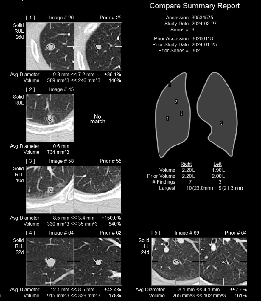
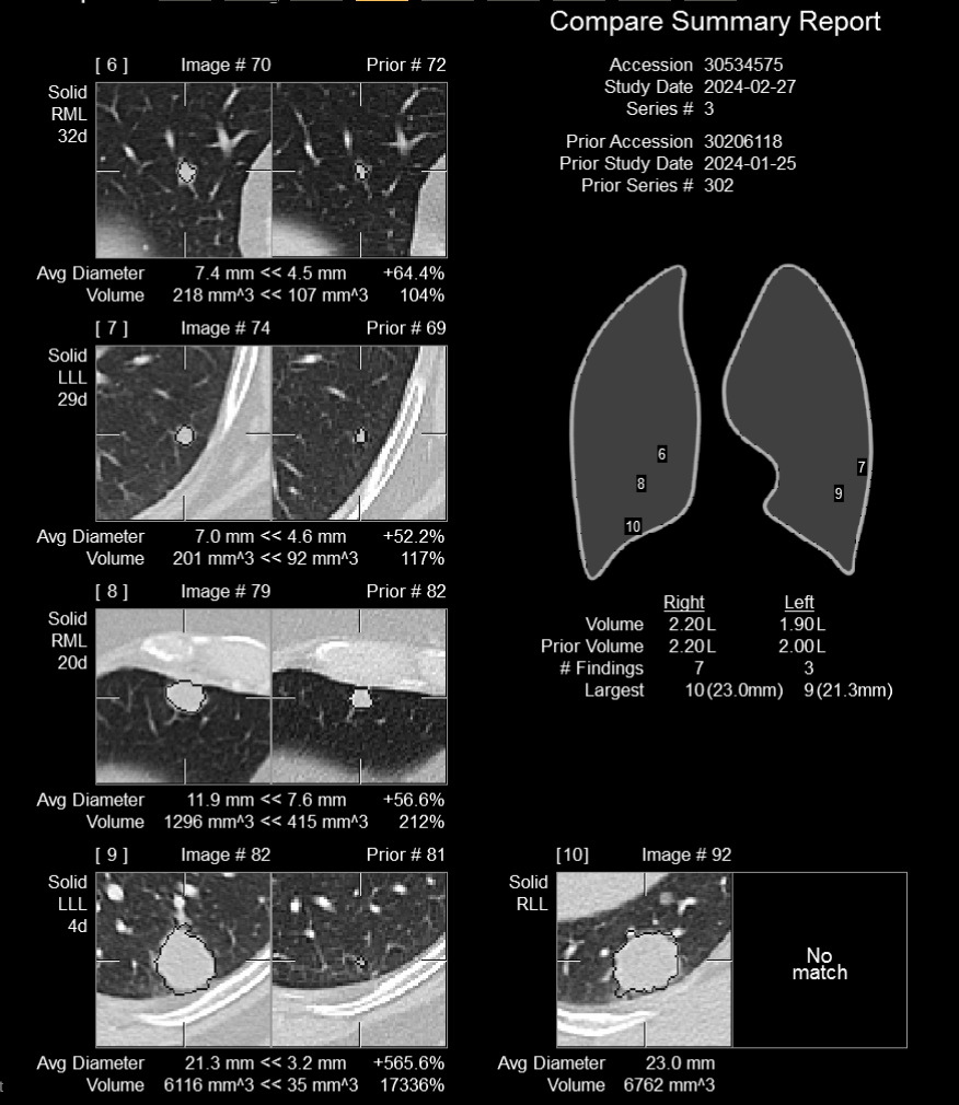
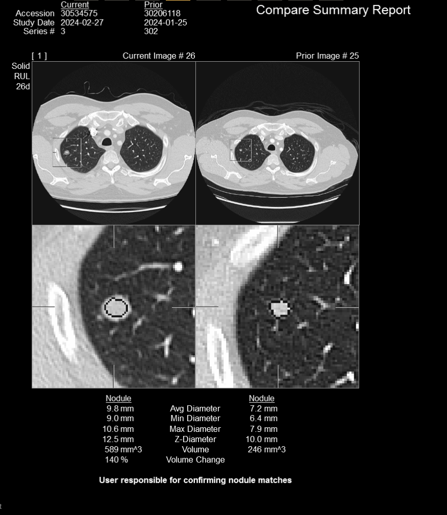
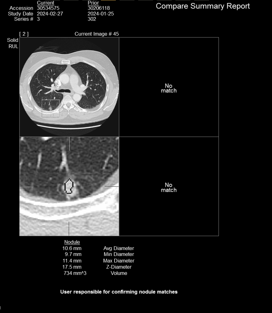
CT Scan Summary:
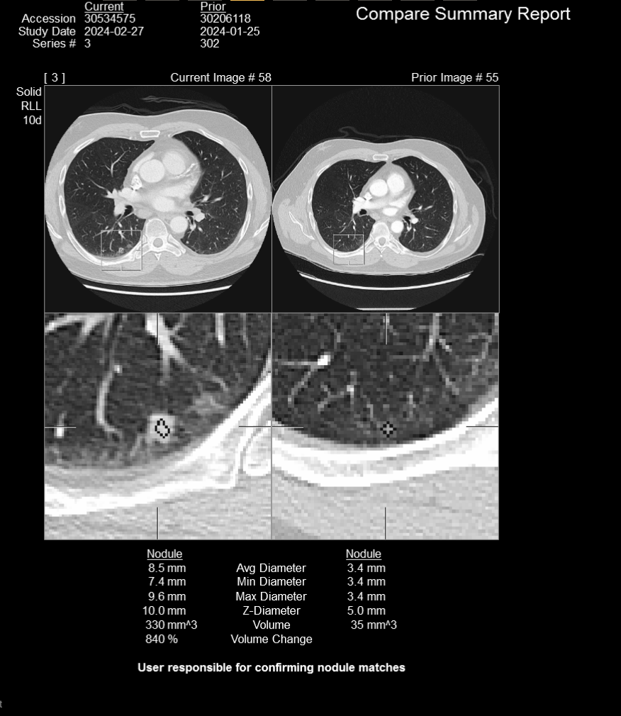
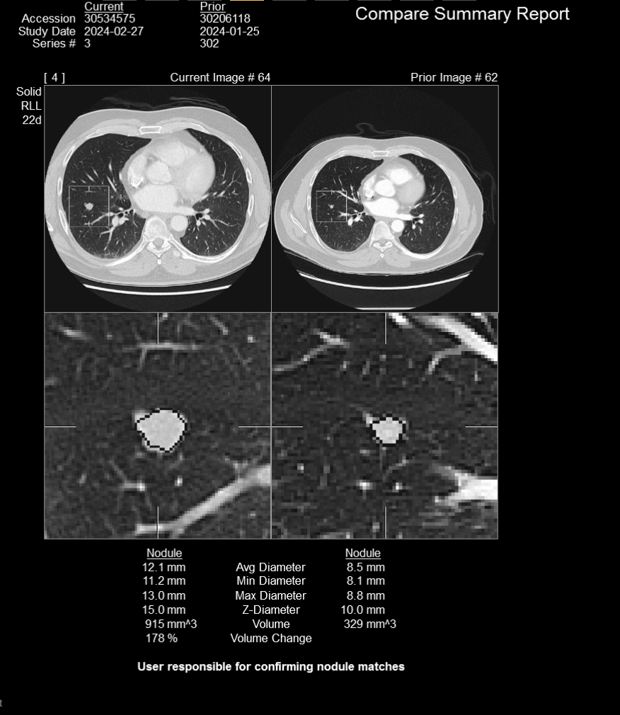
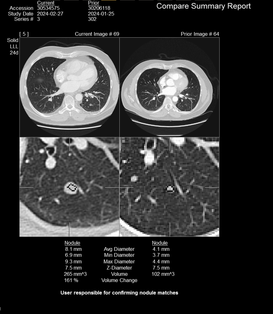
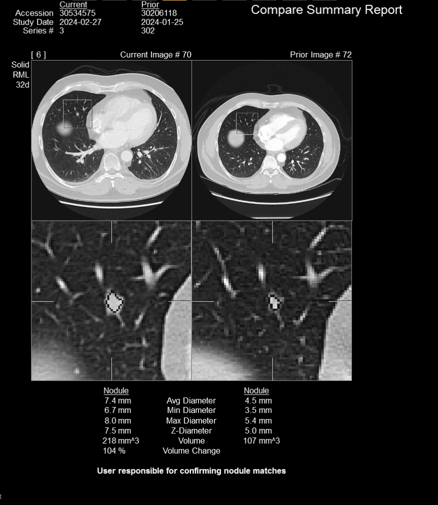
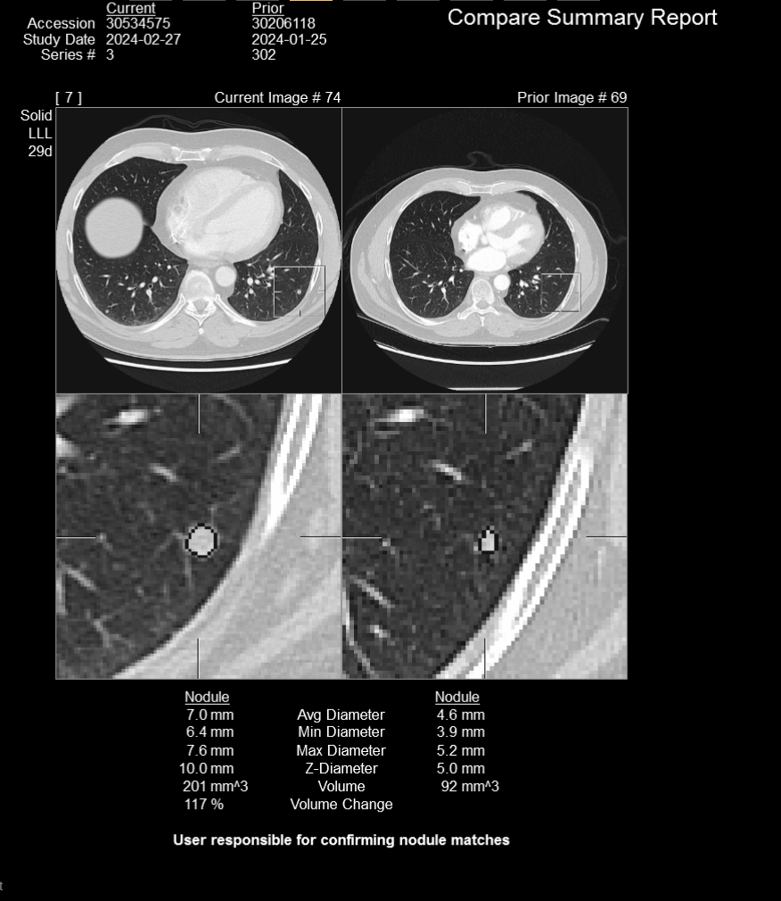
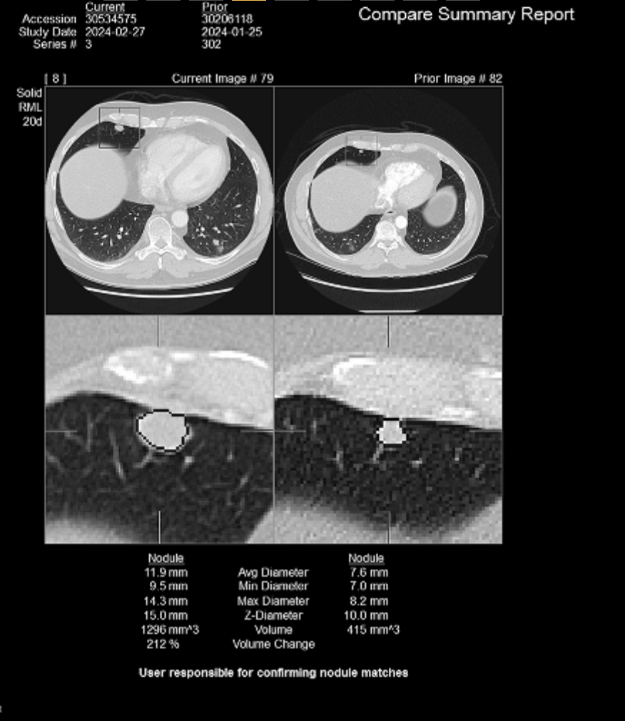
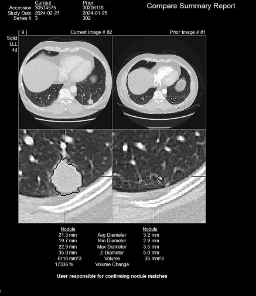
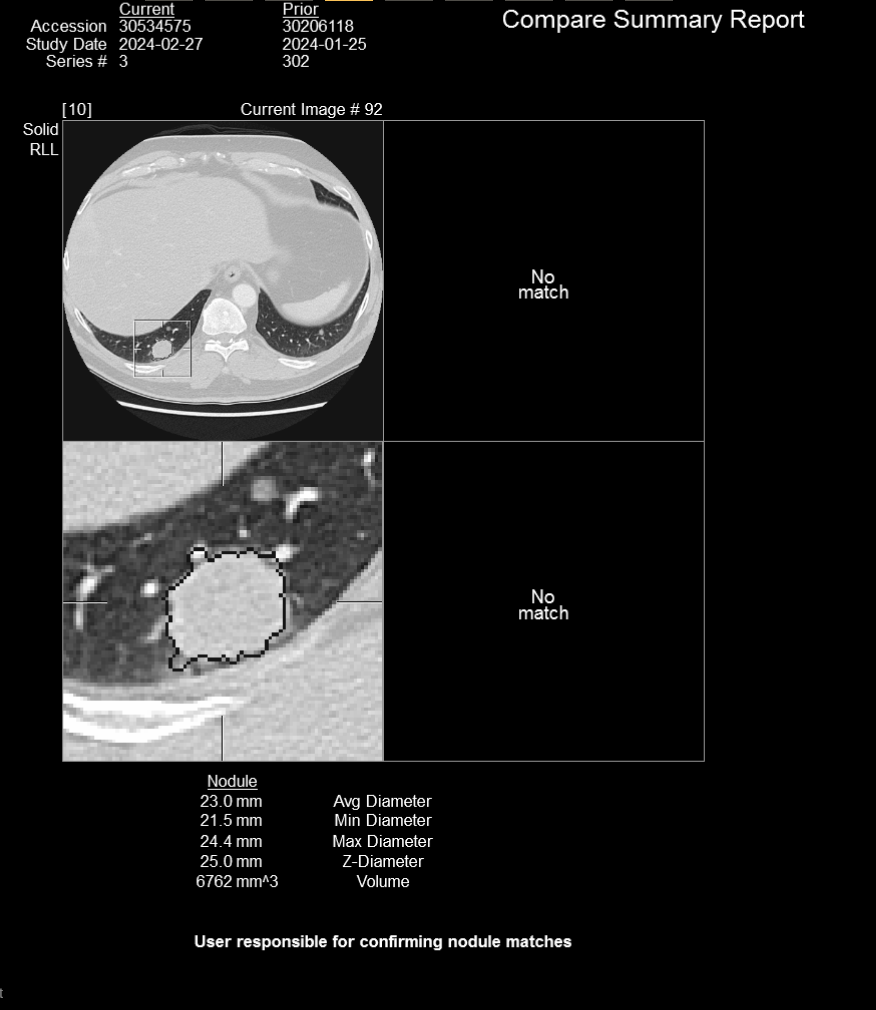
Lungs/Airways/Pleura: Multiple bilateral pulmonary metastases have enlarged. For example, left lower lobe nodule on image 82 of series 3 measures 2.1 cm, previously 1.2 cm. Right upper lobe nodule measures 1.1 cm, previously 0.7 cm, image 26. Left lower lobe nodule measures 0.8 cm, previously 0.5 cm, image 69. Right lower lobe nodule measures 0.9 cm, previously 0.5 cm, image 57. Right lower lobe nodule on image 93 measures 2.4 cm, previously 1.5 cm. The central airways are patent. No pleural effusion.Neck/Mediastinum/Nodes/Heart: Left port catheter terminates in the SVC. Heart size is normal. No pericardial effusion.
Hilar and mediastinal lymph nodes are increased in size. Right hilar lymph node measures 1.9 cm, previously 1.2 cm, image 51 series 2. Subcarinal lymph node measures 1.5 cm, previously 1.1 cm, image 53. Left hilar lymph node measures 1.7 cm, previously 1.2 cm, image 55.
Upper abdomen: Hepatic metastases have enlarged. Left hepatic lobe lesion measures 2.2 cm, previously 1.4 cm, image 102. Right hepatic lobe lesion measures 3.4 cm, previously 2.4 cm, image 97. The remaining visualized upper abdominal structures are unremarkable.
Bones/Soft Tissues: No suspicious skeletal lesions.
With all this “looks terrible on paper” brouhaha I’m still active and in great spirits considering all I’m having to handle.
Here’s the summary of the CT scan from Tuesday - click on any image to open a larger image:
Thank you all for your continued thoughts and prayers.